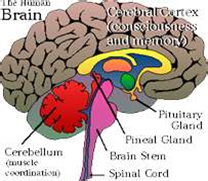
Pages

Selasa, 26 Maret 2013

Pineal Gland


Pineal  gland

    The pineal gland (also called the pineal body or epiphysis) is a small endocrine gland in the brain. It is located near the center of the brain, between the two hemispheres, tucked in a groove where the two rounded thalamic bodies join.it consists of two types of cells 1.parenchymal cells 2.neuroglial cells.

   The pineal gland is a reddish-gray body about the size of a pea (8 mm in humans) located just rostro-dorsal to the superior colliculus and behind and beneath the stria 

   medullaris, between the laterally positioned thalamic bodies. It is part of the epithalamus.
The pineal gland is a midline structure, and is often seen in plain skull X-rays, as it is often calcified.  

The main hormone produced and secreted by the pineal gland is melatonin

Secretion is highest at night and between the ages of 0-5.melatonin acts mainly on gonads.


Sumber : Bpk. Dr. Iskandar Zulkarnain

#posting tugas cyberprenership
    ahmad baihaqi
    NIM 1112503964
 

Sex Organ


Sex organs

The Sex organs (Gonads) are the testes in the male, and the ovaries in the female. Both of these organs produce and secrete hormones that are balanced by the hypothalamus and pituitary glands.
The main hormones from the reproductive organs are:

Testosterone is more prominent in males. It belongs to the family of androgens, which are steroid hormones producing masculine effects. Testosterone stimulates the development and functioning of the primary sex organs. It also stimulates the development and maintenance of secondary male characteristics, such as hair growth on the face and the deep pitch of the voice.
Estrogen In females, this hormone stimulates the development of the uterus and vagina. It is also responsible for the development and maintenance of secondary female characteristics, such as fat distribution throughout the body and the width of the pelvis.


Sumber : Bpk. Dr. Iskandar Zulkarnain

#posting tugas cyberprenership
    ahmad baihaqi
    NIM 1112503964

The pancreas contains exocrine and endocrine cells


The pancreas contains exocrine and endocrine cells.

Groups of endocrine cells, the islets of Langerhans, secrete two hormones. The beta cells secrete insulin; the alpha cells secrete glucagon. The level of sugar in the blood depends on the opposing action of these two hormones.

Insulin decreases the concentration of glucose in the blood. Most of the glucose enters the cells of the liver and skeletal muscles. In these cells, this monosaccharide is converted to the polysaccharide glycogen. Therefore, insulin promotes glycogenesis or glycogen synthesis, in which glucose molecules are added to chains of glycogen. Excess glucose is also stored as fat in adipose tissue cells in response to insulin.

Insulin deficiency leads to the development of diabetes mellitus, specifically type I, juvenile diabetes. As the pancreas does not produce sufficient insulin, it is treated by insulin injections. In type II or maturity onset diabetes, the pancreas does produce enough insulin, but the target cells do not respond to it.


Sumber : Bpk. Dr. Iskandar Zulkarnain

#posting tugas cyberprenership
    ahmad baihaqi
    NIM 1112503964

The pancreas is very important organ in the digestion system


Pancreas

The pancreas is very important organ in the digestion system and the circulatory system because it helps to maintain our blood sugar levels.

The pancreas is considered to be part of the gastrointestinal system. It produces digestive enzymes to be released into the small intestine to aid in reducing food particles to basic elements that can be absorbed by the intestine and used by the body.
 It has another very different function in that it forms insulin, glucagon and other hormones to be sent into the bloodstream to regulate blood sugar levels and other activities throughout the body.

It has a pear-shape to it and is approximately 6 inches long. It is located in the middle and back portion of the abdomen. The pancreas is connected to the first part of the small intestine, the duodenum, and lies behind the stomach. The pancreas is made up of glandular tissue: any substance secreted by the cells of the pancreas will be secreted outside of the organ.


The pancreas is unusual among the body's glands in that it also has a very important endocrine function. Small groups of special cells called islet cells throughout the organ make the hormones of insulin and glucagon. These, of course, are hormones that are critical in regulating blood sugar levels. These hormones are secreted directly into the bloodstream to affect organs all over the body.


Insulin acts to lower blood sugar levels by allowing the sugar to flow into cells. Glucagon acts to raise blood sugar levels by causing glucose to be released into the circulation from its storage sites. Insulin and glucagon act in an opposite but balanced fashion to keep blood sugar levels stable.

    A healthy working pancreas in the human body is important for maintaining good health by preventing malnutrition, and maintaining normal levels of blood sugar. The digestive tract needs the help of the enzymes produced by the pancreas to reduce food particles to their simplest elements, or the nutrients cannot be absorbed. Carbohydrates must be broken down into individual sugar molecules. 
     Proteins must be reduced to simple amino acids. Fats must be broken down into fatty acids. The pancreatic enzymes are important in all these transformations. The basic particles can then easily be transported into the cells that line the intestine, and from there they can be further altered and transported to different tissues in the body as fuel sources and construction materials. Similarly, the body cannot maintain normal blood sugar levels without the balanced action of insulin and glucagon.

Sumber : Bpk. Dr. Iskandar Zulkarnain

#posting tugas cyberprenership
    ahmad baihaqi
    NIM 1112503964

Renin-Angiotensi-Aldosterone system


Adrenal Medulla The hypothalamus starts nerve impulses that travel the path from the bloodstream, spinal cord, and sympathetic nerve fibers to the Adrenal Medulla, which then releases hormones. The effects of these hormones provide a short-term response to stress

Excessive secretion of the glucocorticoids causes Cushing's syndrome, characterized by muscle atrophy or degeneration and hypertension or high blood pressure.
Under secretion of these substances produces Addison's disease, characterized by low blood pressure and stress.

Epinephrine and norepinephrine produce the "fight or flight" response, similar to the effect from the sympathetic nervous system. Therefore, they increase heart rate, breathing rate, blood flow to most skeletal muscles, and the concentration of glucose in the blood. They decrease blood flow to the digestive organs and diminish most digestive processes.


The adrenal medulla secretes two hormone, adrenalin or epinephrine and noradrenalin or norepinephrine, whose functions are very similar but not identical. The adrenal medulla is derived embriogically from neural tissue. It has been likened to an overgrown sympathetic ganglion whose cell bodies do not send out nerve fibers, but release their active substances directly into the blood, thereby fulfilling the criteria for an endocrine gland. In controlling epinephrine secretion, the adrenal medulla behaves just like any sympathetic ganglion, and is dependent upon stimulation by sympathetic preganglionic fibers.

Epinephrine promotes several responses, all of which are helpful in coping with emergencies: the blood pressure rises, the heart rate increases, the glucose content of the blood rises because of glycogen breakdown, the spleen contracts and squeezes out a reserve supply of blood, the clotting time decreases, the pupils dilate, the blood flow to skeletal muscles increase, the blood supply to intestinal smooth muscle decreases and hairs become erect.  
These adrenal functions, which mobilize the resources of the body in emergencies, have been called the fight-or-flight response. Norepinephrine stimulates reactions similar to those produced by epinephrine, but is less effective in conversion of glycogen to glucose.


fight-or-flight response


Sumber : Bpk. Dr. Iskandar Zulkarnain

#posting tugas cyberprenership
    ahmad baihaqi
    NIM 1112503964


Adrenal glands are a pair of ductless


Adrenal glands

Adrenal glands are a pair of ductless glands located above the kidneys. Through hormonal secretions, the adrenal glands regulate many essential functions in the body, including biochemical balances that influence athletic training and general stress response.

The glucocorticoids include corticosterone, cortisone, and hydrocortisone or cortisol. These hormones serve to stimulate the conversion of amino acids into carbohydrates which is a process known as gluconeogenesis, and the formation of glycogen by the liver.

They also stimulate the formation of reserve glycogen in the tissues, such as in the muscles. The glucocorticoids also participate in lipid and protein metabolism. The cortex of the adrenal gland is known to produce over 20 hormones, but their study can be simplified by classifying them into three categories: glucocorticoids, mineralcorticoids, and sex hormones.

They are triangular-shaped glands located on top of the kidneys. They produce hormones such as estrogen, progesterone, steroids, cortisol, and cortisone, and chemicals such as adrenalin (epinephrine), norepinephrine, and dopamine. When the glands produce more or less hormones than required by the body, disease conditions may occur.

    The adrenal cortex secretes at least two families of hormones, the glucocorticoids and mineral corticoids. The adrenal medulla secretes the hormones epinephrine (adrenalin) and norepinephrine (noradrenalin).

Adrenal Cortex: 

     The hormones made by the Adrenal Cortex supply long-term responses to stress. The two major hormones produced are the Mineral Corticoids and the Glucocorticoids. The Mineral Corticoids regulate the salt and water balance, leading to the increase of blood volume and blood pressure.
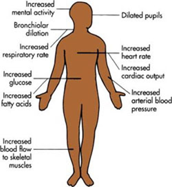
The Glucocorticoids are monitoring the ACTH, in turn regulating carbohydrates, proteins, and fat metabolism. This causes an increase in blood glucose. Glucocorticoids also reduce the body's inflammatory response.

    Cortisol is one of the most active glucocorticoids. It usually reduces the effects of inflammation or swelling throughout the body. It also stimulates the production of glucose from fats and proteins, which is a process referred to as gluconeogenesis.

    Aldosterone is one example of a mineralcorticoid. It signals the tubules in the kidney nephrons to reabsorb sodium while secreting or eliminating potassium. If sodium levels are low in the blood, the kidney secretes more renin, which is an enzyme that stimulates the formation of angiotensin from a molecule made from the liver. Angiotensin stimulates aldosterone secretion. As a result, more sodium is reabsorbed as it enters the blood.

    Aldosterone, the major mineralcorticoid, stimulates the cells of the distal convoluted tubules of the kidneys to decrease re-absorption of potassium and increase re-absorption of sodium. This in turn leads to an increased re-absorption of chloride and water. These hormones, together with such hormones as insulin and glucagon, are important regulators of the ionic environment of the internal fluid.

    The renin-angiotensin-aldosterone mechanism can raise blood pressure if it tends to drop. It does this in two ways. Angiotensin is a vasoconstrictor, decreasing the diameter of blood vessels. As vessels constrict, blood pressure increases. In addition, as sodium is reabsorbed, the blood passing through the kidney becomes more hypertonic. Water follows the sodium into the hypertonic blood by osmosis. This increases the amount of volume in the blood and also increases the blood pressure.


Sumber : Bpk. Dr. Iskandar Zulkarnain

#posting tugas cyberprenership
    ahmad baihaqi
    NIM 1112503964


Senin, 25 Maret 2013

The parathyroid hormone


The parathyroid hormone, called parathormone, regulates the calcium-phosphate balance between the blood and other tissues.

Production of this hormone is directly controlled by the calcium concentration of the extracellular fluid bathing the cells of these glands.

Parathormone exerts at least the following five effects:
 
(1)it increases gastrointestinal absorption of calcium by stimulating the active transport system and moves calcium from the gut lumen into the blood;
(2) it increases the movement of calcium and phosphate from bone into extracellular fluid. This is accomplished by stimulating osteoclasts to break down bone structure, thus liberating calcium phosphate into the blood. In this way, the store of calcium contained in bone is tapped;
(3)it increases re-absorption of calcium by the renal tubules, thereby decreasing urinary calcium excretion;
(4)it reduces the re-absorption of phosphate by the renal tubules
(5)it stimulates the synthesisof 1,25-dihydrixycholecalciferol by the kidney.
 
        
If parathyroid glands are removed accidentally during surgery on the thyroid, there would be arise in the phosphate concentration in the blood.

There would also be a drop in the calcium concentration as more calcium is excreted by the kidneys and intestines, and more incorporated into the bone.

This can produce serious disturbances, particularly in the muscles and nerves, which use calcium ions for normal functioning.

Over activity of the parathyroid glands, which can result from a tumor on the glands, produces a weakening of the bones.

This is a condition that makes them much more vulnerable to fracturing because of excessive withdrawal of calcium from the bones.


Sumber : Bpk. Dr. Iskandar Zulkarnain

#posting tugas cyberprenership
    ahmad baihaqi
    NIM 1112503964


Parathyroid Gland


Parathyroid gland
   
    There are four parathyroid glands. They are small, light-colored lumps that stick out from the surface of the thyroid gland. All four glands are located on the thyroid gland.
They are butterfly-shaped and located inside the neck, more specifically on both sides of the windpipe.

   One of the parathyroid glands most important functions is to regulate the body's calcium and phosphorus levels. Another function of the parathyroid glands is to secrete parathyroid hormone, which causes the release of the calcium present in bone to extracellular fluid.
PTH does this by depressing the production of osteoblasts, special cells of the body involved in the production of bone and activating osteoclasts, other specialized cells involved in the removal of bone.


Receptors bind a specific hormone and the result is a specific physiologic response, meaning a normal response of the body.

    PTH finds its major target cells in bone, kidneys, and the gastrointestinal system.
Calcitonin, a hormone produced by the thyroid gland that also regulates ECF calcium levels and serves to counteract the calcium-producing effects of PTH.

    The adult body contains as much as 1 kg of calcium. Most of this calcium is found in bone and teeth. The four parathyroid glands secrete the parathyroid hormone (PTH). It opposes the effect of thyrocalcitonin. It does this by removing calcium from its storage sites in bones, releasing it into the bloodstream. It also signals the kidneys to reabsorb more of this mineral, transporting it into the blood. It also signals the small intestine to absorb more of this mineral, transporting it from the diet into the blood.

    Calcium is important for steps of body metabolism. Blood cannot clot without sufficient calcium. Skeletal muscles require this mineral in order to contract. A deficiency of PTH can lead to tetany, muscle weakness due to lack of available calcium in the blood.


Sumber : Bpk. Dr. Iskandar Zulkarnain

#posting tugas cyberprenership
    ahmad baihaqi
    NIM 1112503964


Thyroid System


    Iodine deficient goiter, (enlargement of the thyroid gland) results because iodine is a crucial component of active thyroid hormones. If there is a low level of iodine in the diet, then less active T3 and T4 can be synthesized. As diagrammed in the figure, when there is less T3 and T4, there is reduced negative feedback inhibition on secretion of the tropic hormones, TRH (thyrotropin releasing hormone; released by the hypothalamus) and TSH (thyroid stimulating hormone or thyrotropin; released by the anterior pituitary). TSH stimulates all aspects of thyroid hormone synthesis; it also stimulates proliferation of follicle cells. When iodine in the diet is low but not too low, individuals may have goiter and yet be euthyroid, because the enlarged thyroid gland is better able to use the limited amount of iodine available. This is an example of hormonal homeostasis.


    Graves disease is the most common cause of hyperthyroidism. Graves disease is an autoimmune disorder in which antibodies are produced that bind to the TSH receptor. Instead of destroying the receptor, these antibodies activate it, stimulating synthesis of T3 and T4, and causing growth of the thyroid gland. Negative feedback inhibition is increased, so the levels of TRH and TSH decrease, but this does not decrease thyroid hormone production because the stimulation of the thyroid gland is independent of TSH. Measurement of low TSH levels is a key diagnostic test for hyperthyroidism


Sumber : Bpk. Dr. Iskandar Zulkarnain

#posting tugas cyberprenership
    ahmad baihaqi
    NIM 1112503964


The Thyroid gland


   The Thyroid gland

     The Thyroid gland is one of the largest endocrine glands in the body. It is positioned on the neck just below the Larynx and has two lobes with one on either side of the trachea. It is involved in the production of the hormones T3 (triiodothyronine) and T4 (thyroxine). These hormones increase the metabolic activity of the body‘s cells. The thyroid also produces and releases the hormone calcitonin (thyrocalcitonin) which contributes to the regulation of blood calcium levels. Thyrocalcitonin or calcitonin decreases the concentration of calcium in the blood. Most of the calcium removed from the blood is stored in the bones.

   The thyroid hormone consists of two components, thyroxine and iodine. This hormone increases the metabolism of most body cells. A deficiency of iodine in the diet leads to the enlargement of the thyroid gland, known as a simple goiter. Hypothyroidism during early development leads to cretinism. In adults, it produces myxedema, characterized by obesity and lethargy. Hyperthyroidism leads to a condition known as exophthalmic goiter, characterized by weight loss as well as hyperactive and irritable behavior.

The thyroid gland is a two-lobed gland that manifests a remarkably powerful active transport mechanism for up-taking iodide ions from the blood.

    As blood flows through the gland, iodide is converted to an active form of iodine. This iodine combines with an amino acid called tyrosine. Two molecules of iodinated tyrosine then combine to form thryroxine. Following its formation, the thyroxine becomes bound to a polysaccharide-protein material called thyroglobulin. The normal thyroid gland may store several weeks supply of thyroxine in this bound form. An enzymatic splitting of the thyroxine from the thyroglobulin occurs when a specific hormone is released into the blood.

   This hormone, produced by the pituitary gland, is known as thyroid-stimulating hormone (TSH). TSH stimulates certain major rate-limiting steps in thyroxine secretion, and thereby alters its rate of release. A variety of bodily defects, either dietary, hereditary, or disease induced, may decrease the amount of thyroxine released into the blood.


   Thyroxine serves to stimulate oxidative metabolism in cells; it increases the oxygen consumption and heat production of most body tissues, a notable exception being the brain. Thyroxine is also necessary for normal growth. The most likely explanation being that thyroxine promotes the effects of growth hormone on protein synthesis. The absence of thyroxine significantly reduces the ability of growth hormone to stimulate amino acid uptake and RNA synthesis. Thyroxine also plays a crucial role in the closely related area of organ development, particularly that of the central nervous system.

   If there is an insufficient amount of thyroxine, a condition referred to as hypothyroidism results. Symptoms of hypothyroidism stem from the fact that there is a reduction in the rate of oxidative energy-releasing reactions within the body cells. Usually the patient shows puffy skin, sluggishness, and lowered vitality. Other symptoms of hypothyroidism include weight gain, decreased libido, inability to tolerate cold, muscle pain and spasm, insomnia and brittle nails. 


Sumber : Bpk. Dr. Iskandar Zulkarnain

#posting tugas cyberprenership
    ahmad baihaqi
    NIM 1112503964
tanahsakti SUDUT KANAN ATAS